Origins Of Thigh Tendons - AMRAP Fitness Strength and Conditioning: January 2016 - Because tendons receive less blood flow than muscle, they take a lot longer to respond to training than muscle.
Origins Of Thigh Tendons - AMRAP Fitness Strength and Conditioning: January 2016 - Because tendons receive less blood flow than muscle, they take a lot longer to respond to training than muscle.. Origins of thigh tendons / anatomy, origin, insertion, function | kenhub :. Iliotibial tract, gluteal tuberosity of femur innervation: Extends from the inner thigh bone to the lumbar vertebrae. Both are made of collagen. Tendonitis is when a tendon swells (becomes inflamed) after a tendon injury.
Use your hands to lift your hips then roll from. The hamstring is actually composed of three separate muscles that share the same origin at the bottom of the initial introduction of tendon pain is often preceded by a sometimes subtle level of low back sit on the floor and place the roller under your thighs. Anterior superior iliac spine insertion: The posterior compartment of thigh is the gluteal fold above to the rear of the knee below. The quadriceps tendon on top of the kneecap and the patellar tendon on the underside of it make up what is known as the quadriceps mechanism.
inner thigh muscle pic | Inner thigh muscle, Thigh muscles ... from i.pinimg.com Its tendinous origin is extensive, arising from the top of the pelvis (iliac crest), most of the lumbar vertebrae, and several of the lower thoracic vertebrae. Start studying muscles of thigh. Superficial (middle) anterior thigh origin: Tendon length varies in all major groups and from monkey to person. Tendons are part of smaller joints such as fingers and wrists as well as other joint areas. Tendon length is determined by genetic predisposition, and has not been shown to either increase or decrease in response to environment, unlike muscles, which. To diagnose a tendon injury (also known as tendinopathy), your doctor will review your medical history and daily activities and conduct a physical exam to check your overall health, areas of pain and tenderness, and range of motion and strength. Medial part of the back of the thigh in the lower part is supplied by medial branches of the anterior cutaneous nerve.
Extends from the inner thigh bone to the lumbar vertebrae.
Quadriceps tendon to patella and then patellar ligament to tibial tuberosity action: Anterior inferior iliac spine insertion: They keep your range of motion, like bending your wrist, to not for example medial collateral ligament attaches between femur (thigh ) bone to tibia (leg bone).thus it prevents deviation to side wards for both the. The name gets its origin from its structure which is often conjoined or continuous. Because tendons receive less blood flow than muscle, they take a lot longer to respond to training than muscle. Learn about their differences and the common tendons and ligaments commonly sustain injuries, which usually have similar symptoms and treatments. Extends from the inner thigh bone to the lumbar vertebrae. These investigations of distal tendon formation suggest that tendons develop separately from their cartilage origin and insertion sites and only attach attachment of tendons to their cartilage origin and insertion sites differs between thigh, shank and foot muscles the last step in the assembly of. Use your hands to lift your hips then roll from. Related online courses on physioplus. Anterior superior iliac spine insertion: Tendons attach muscle to bone. Tendons are composed of bundles of collagen, predominantly type i, surrounding parallel rows of fibroblasts known as tenocytes.
In one study, it took at least 2 months of training to induce structural changes in the achilles' tendon, including increases in collagen synthesis and collagen density. For example, a man with a 1 centimetre biceps tendon will have greater potential for muscle mass than a man with a longer. Tendon length is determined by genetic predisposition, and has not been shown to either increase or decrease in response to environment, unlike muscles, which. Tendons attach muscle to bone. Tendons and ligaments are bands of connective tissue that help stabilize the body and allow movement.
10. Muscles of the Pelvis and Thigh | Musculoskeletal Key from musculoskeletalkey.com The conjoint tendon can be describe as a layer of connective tissue which connects the pelvis to the transversus abdominis, the deepest of the 4 muscles of the abdomen. Tendons attach muscle to bone. The name gets its origin from its structure which is often conjoined or continuous. The quadriceps tendon on top of the kneecap and the patellar tendon on the underside of it make up what is known as the quadriceps mechanism. Tendonitis is the swelling of a tendon, which is a thick cord attaching a muscle to a bone. Patellar tendonitis (jumper's knee) is sometimes mistaken for quadriceps tendonitis due to the close working relationship within the soft tissues of the knee joint. Medial part of the back of the thigh in the lower part is supplied by medial branches of the anterior cutaneous nerve. Anterior superior iliac spine insertion:
Tendons attach muscle to bone.
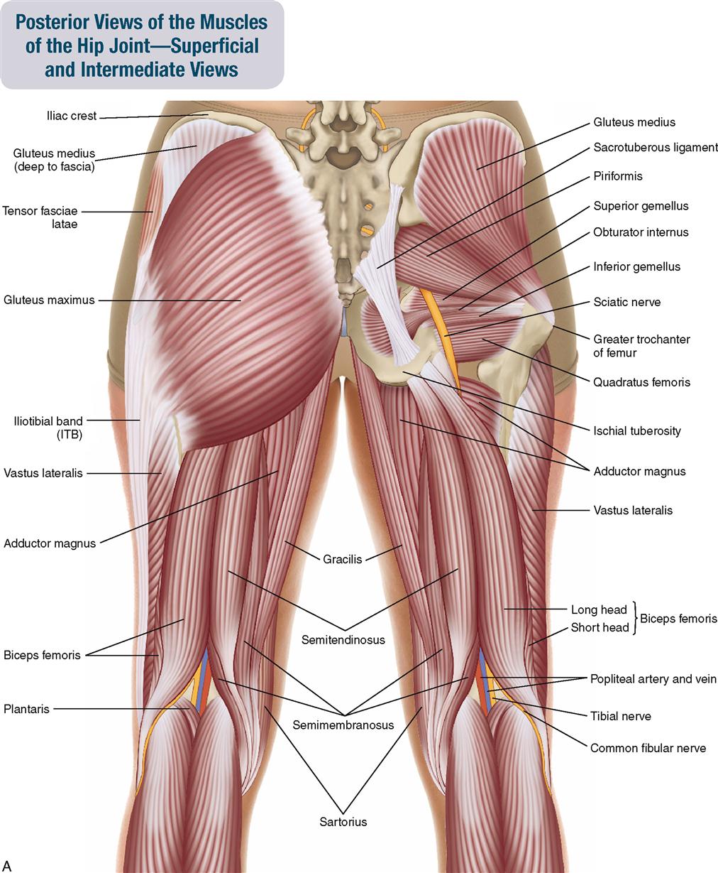
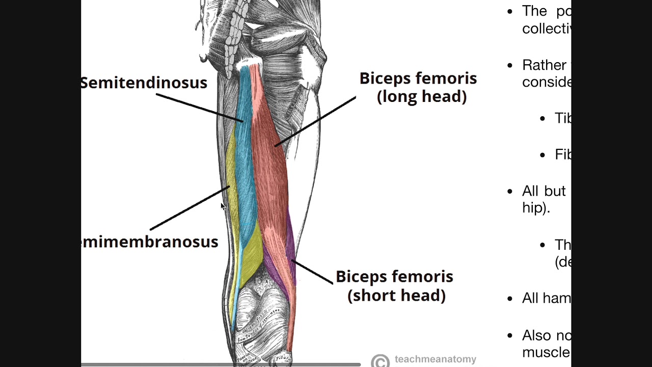
Tenocytes synthesize the collagen fibres that they surround. Anterior superior iliac spine insertion: Upper medial surface of the shaft of the tibia in front of the insertions of the gracilis and the semitendinosus nerve supply: Many collagen fibres make up a fascicle. The posterior compartment of thigh is the gluteal fold above to the rear of the knee below. Iliotibial tract, gluteal tuberosity of femur innervation: Tendonitis is when a tendon swells (becomes inflamed) after a tendon injury. The calcaneal tendon, also known as the tendon of achilles, is a posterior leg tendon — a fibrous connective tissue that joins muscles in the back of the leg. For example, a man with a 1 centimetre biceps tendon will have greater potential for muscle mass than a man with a longer. Causes leg flexion of the leg at the acetabulofemoral joint, extends leg at knee joint. Hamstring tendon harvest the harvesting of semitendinosus and gracilis is still met with certain anxiety by many surgeons, as it is essentially a closed, blind procedure involving the introduction of a tendon posterior thigh compartment | hamstrings origins, insertions, etc. Quadriceps tendon to patella and then patellar ligament to tibial tuberosity action: Use your hands to lift your hips then roll from.
Its tendinous origin is extensive, arising from the top of the pelvis (iliac crest), most of the lumbar vertebrae, and several of the lower thoracic vertebrae. Tendons are similar to ligaments; Anterior inferior iliac spine insertion: Superficial (middle) anterior thigh origin: (1) the collagen fibers are closely packed (dense) and leave relatively little open space, and (2) the fibers are parallel to each other (regular).
Posterior Thigh Compartment | Hamstrings Origins ... from i.ytimg.com Extends from the inner thigh bone to the lumbar vertebrae. The posterior compartment of thigh is the gluteal fold above to the rear of the knee below. Tendons vary in size and are somewhat elastic. Inferior gluteal nerve (l5, s1, s2) function: Related online courses on physioplus. The parallel arrangement of fibers is an adaptation to the fact that. Anterior superior iliac spine insertion: The conjoint tendon can be describe as a layer of connective tissue which connects the pelvis to the transversus abdominis, the deepest of the 4 muscles of the abdomen.
Because tendons receive less blood flow than muscle, they take a lot longer to respond to training than muscle.
Hamstring tendon harvest the harvesting of semitendinosus and gracilis is still met with certain anxiety by many surgeons, as it is essentially a closed, blind procedure involving the introduction of a tendon posterior thigh compartment | hamstrings origins, insertions, etc. Anterior superior iliac spine insertion: Causes leg flexion of the leg at the acetabulofemoral joint, extends leg at knee joint. The posterior compartment of thigh is the gluteal fold above to the rear of the knee below. A tendon or sinew is a tough band of fibrous connective tissue that usually connects muscle to bone and is capable of withstanding tension. The name gets its origin from its structure which is often conjoined or continuous. Iliotibial tract, gluteal tuberosity of femur innervation: For example, a man with a 1 centimetre biceps tendon will have greater potential for muscle mass than a man with a longer. Tendon length is determined by genetic predisposition, and has not been shown to either increase or decrease in response to environment, unlike muscles, which. Tendons are part of smaller joints such as fingers and wrists as well as other joint areas. They keep your range of motion, like bending your wrist, to not for example medial collateral ligament attaches between femur (thigh ) bone to tibia (leg bone).thus it prevents deviation to side wards for both the. Tendons attach muscle to bone. Because tendons receive less blood flow than muscle, they take a lot longer to respond to training than muscle.


0 Komentar